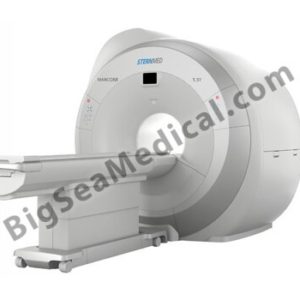
|
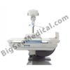
Technical data Xenox DR1000F
|
Sternmed Digital Fluoroscopy and Radiography System
|
|
Multifunctional Remote Control Table
|
|
|
Control Mode
|
Compartment remote control、Bed-close control;automatic identification,slow startslow stop
|
|
Table Rotating Motion
|
Rotating Range 0 °~+90° and 0 °~-25°
|
|
Rotating Speed 90 °/22s
|
|
Table Transverse Moving
|
Moving Range(horizontal)is 220mm
|
|
Moving Speed(horizontal)is 220mm/10s
|
|
Image System Moving
|
Moving Range is 740mm when table rotating angel is -12 °~+90 °,and it is 340mm when table rotating angel is -12 °~-25 °
|
|
Moving speed is 740mm/22s when the surface of the table is horizontal
|
|
Tube Moving Device
|
The Distance from focus to film is1100mm; could be extend to 1500mm when it is vertical position; It has two modes which are 1100mm、1500mm,manual adjustment
|
|
Compression Device
|
Straight line moving range(compress head face to table)is 100mm
|
|
Moving time from rollback position to forward extreme position is 4~7s
|
|
Compress power is 78~98N(8~10kgf)when table is horizontal
|
|
Compress power is 69~98N(7~10kgf) when table surface is vertical position
|
|
High Voltage Generator: EMD
|
|
kV Range
|
40-150kV(step 1kV)
|
|
mA Range
|
10mA~630mA(adjustable)
|
|
Nominal Power
|
50kW
|
|
Exposure Time
|
0.001s-10s
|
|
Inverter Frequency
|
240kHz
|
|
Maximum kV
|
150kV
|
|
mAs Range(mAs)
|
0.4~1000mAs
|
|
Fluoroscopy kV Range
|
40~125(step 1kV)
|
|
Fluoroscopy mA Range(mA)
|
0.1~10mA
|
|
Maximum alarm time for fluoroscopy
|
5min
|
|
Preset Exposure Parameter
|
2000
|
|
X-RayTube: Varian
|
|
Power
|
32/77kW
|
|
Focus
|
0.6mm/1.2mm
|
|
Maximum kV
|
150kV
|
|
Anode Heat Capacity
|
300kHU
|
|
Anode Rotation Speed
|
8500rpm
|
|
Grid
|
|
Grid Focal Length
|
110cm
|
|
Grid Ratio
|
10:1
|
|
Grid Density
|
103 LP/Inch (40LP/cm)
|
|
Grid Size
|
15″×18″
|
|
Image Intensifier: Thales
|
|
Screen Size
|
9″
|
|
Center Area Resolution
|
46LP/cm
|
|
CCD Camera
|
|
Sampling Resolution
|
1k× 1k × 12bit
|
|
Sampling Gray Scale
|
12bit
|
|
Fluoroscopy Sampling Rate
|
23 frame / s
|
|
Subtration Speed
|
7.5 frame / s
|
|
Digital Spot Film
|
7.5 frame / s
|
|
Image Noise Reduction
|
Real time noise reduction、Dynamic detection
|
|
FPD Detector:Varian
|
|
Imaging Area
|
14×17 inch
|
|
Imaging Medium
|
A-Si (Amorphous Silicon)
|
|
Preview Time
|
About 3s
|
|
A/D Output
|
14bits
|
|
Pixel Matrix
|
3072 x 2560
|
|
Pixel Size
|
139 μm
|
|
Spatial Resolution
|
3.6lp/mm
|
|
Software System Function
|
|
Local Register Patients ‘Information
|
Register examination number of patient、Patient Number、Name、Gender、Age、Birthday、Examine description
|
|
Obtain Patients’ Information Through Worklist
|
Refresh Patient List;Select inquire condition、Input inquire condition、Find patient through inquire condition
|
|
Emergency Treatment
|
To critical patient it could directly get into examine without register
|
|
Patient Information revise/delete
|
Revise and delete the registered information of the patient
|
|
Historical Examination of The Patient
|
Re-examination the historical examination patient、inquire、data-transmit、patient information delete
|
|
Gastrointestinal Software Collection Processing Function
|
Image Analysis、Adjust、Storage、Freeze(fluoroscopy the last one),Adjust window width and position,Real time noise reduction,Image enhancement,Real time digital subtration、Post-processing subtration、Image positive and negative transfer;Image vertical、flip horizontal、90 ° Rotation、Electronic Shearing、Image sharpening、Zoom、Gray Scale stretching、Mark、Measure、Multiple image display、Digital replay,Patients’ profile image repoty printout、Storage、CD Burner
|
|
DR Software Post-processing
|
Adjust window width and position,Image enhancement,Image vertical、flip horizontal、90 ° Rotation、Cut out、Image sharpening、Zoom、Gray Scale stretching、Mark、Multiple image display、Digital
|
|
Image Transmit
|
Image Transmit Dicom Store
|
|
Image Print
|
Transmit image to printer、need to have peint preview layout function、print settings(priority、destination、medium type、magnify type、smoothness、border density、space density)
|

![xenox-dr1000f[1] xenox-dr1000f[1]](https://www.bigseamedical.com/wp-content/uploads/2015/06/xenox-dr1000f1.jpg)
