Product Description
|
Technical data Marcom 1.5T |
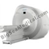
Sternmed Superconductive 1.5T MRI |
|
Superconductive Magnet |
|
|
Superconductive Magnet |
1.5T active shield superconducting magnet |
|
Homogeneity |
≤5.0 ppm @ DSV 45cm ≤1.0 ppm @ DSV 40cm |
|
Stability |
≤0.02 ppm/h |
|
Helium boil-off rate |
<50 cc/h |
|
Helium refill period |
≥24 months |
|
5GS (X,Y,Z) |
≤2.5m,2.5m,4m |
|
Patient aperture |
600mm |
|
Gradient |
|
|
Gradient System |
Active shield full body gradient coil systems, noise reduction & mute technology |
|
Gradient Coil |
England Telsa Corporation |
|
Cooling System Type |
Water-cooled |
|
Cold Head |
Sumitomo cold head |
|
Gradient strength |
Single axis 34mT/m, Effective 59 mT/m |
|
Slew rate |
Single axis 130mT/m/ms, Effective 225mT/m/ms |
|
RF System |
|
|
Spectrometer |
All-digital (8 channnels) |
|
Power of transmitter amplifier |
18KW |
|
Receiving coils |
Built-in preamplifier phased array coil |
|
Receiving coil type (standard) |
head, neck, body, knee, spine, shoulders, Breast coil |
|
Workstation |
|
|
Operating system |
Windows XP Professional |
|
CPU |
Dual frequency ≥2.8GHz |
|
RAM |
≥2GB |
|
Hard disk |
≥300GB |
|
The main screen displays |
≥22´´LCD TFT monitor |
|
Screen monitoring device, machine monitoring, patient monitoring, multi-parameter monitor the amount of concentrated, high security |
|
|
Machine control parameters |
pressure, level, temperature |
|
Patient monitoring parameters |
electrocardio, respiratory, RF, db/dt |
|
System operating modes |
normal operation mode, a controlled operating mode |
|
Image processing and analysis |
image enhancement, zoom, pan, crop, negatives, window width and window level adjustments, mark or clear text, distance measurement, regions of interest selected, the average pixel value, standard deviation of pixel values, signal values distribution, projection reconstruction |
|
Network components |
DICOM 3.0 standard interface, through the local Ethernet network easily to link camera, diagnosis and treatment workstations, medical information systems, remote diagnostics system. |
|
Pulse sequences |
|
|
|
2D, 3D GRE 2D, 3D SE 2D, 3D FSE FSPGR(positive/ negative phase sequence) TrueFISP(Fast steady precession fast imaging) SSPGRE(Steady state process gradient echo) FRFSE(Fast recovery fast spin echo) Single-shot FSE 2D (Single shot fast spin echo) MSFSE(Multi shot fast spin echo) FSE 2D with Inversion Recovery (2D reversion fast spin echo sequence) STIR(Short time inversion recovery) FLAIR(Fluid attenuated inversion recovery) MRM,MRU,MRCP EPI SE-based EPI SE-based DW DWI(Diffusion weighted imaging) 2D, 3D MRA Automatic pre-scan |
|
Gate Trigger |
Breathing, electrocardio |
|
Reconstruction algorithm |
1DFFT, 2DFFD, 2DFFT, MIP, MPR, SSD |
|
Scanning parameter |
|
|
Resolution |
0.75mm, Head,24cmFOV 256×256 1.0mm, Body, 30cmFOV 256×256 0.5mm, Head , 24cmFOV 512×512 0.75mm, Body,30cmFOV 512×512 |
|
Acquisition matrix |
64/128/ 256/ 512/1024 |
|
Maximum display matrix |
Maximum display matrix1024x1024 |
|
FOV |
10~450 mm |
|
Scan orientations |
Any angle (axial, sagittal, coronal, any slope, multi-layer multi-angle) |
|
Image type |
T1 weighted imaging, T2 weighted imaging, T2*weighted imaging, proton density imaging, Water suppressed imaging, Fat Suppressed imagine, MRM, MRU, MRCP, Magnetic Resonance angiography(MRA), Diffusion weighted imaging(DWI) |
|
Patient Table |
|
|
Patient Table |
Drop out of the open two-dimensional movement, motor drives, cross laser positioning, Emergency braking situation or power outage, you can manually take the bed. |
|
Status display and Configuration |
8” TFT- LCD display, resolution 800 * 600, real-time display system status |
|
Max. Patient Load |
200Kg |
|
Positioning accuracy |
<1mm |
|
Operating position |
Bilateral |
|
Positioning Accessories |
mattress, pillow, head pillow, various parts of the fixed pad |
|
Power supply |
|
|
Helium compressor |
3N-a.c 380~415V/50Hz,6.9KW |
|
Machine |
3N-a.c.380V±38V/50Hz±1Hz,50KW q |
|
Typical Layout |
|
|
Magnet Room |
About 33m2(5m×6.5m); |
|
Equipment Room |
About 24m2(3m×8m); |
|
Control Room |
About 7.5m2(1.5m×5m); |
|
Total MRI-System area |
About 64.5m2. |

![marcom-1-5t[1] marcom-1-5t[1]](https://www.bigseamedical.com/wp-content/uploads/2015/06/marcom-1-5t1.jpg)
